Researchers use slide scanning microscopy to investigate CD8 T cells in pancreatic tissue
Dr. Matthias von Herrath is a professor of autoimmunity and inflammation at the La Jolla Institute for Immunology (USA). His lab is committed to the understanding of type 1 diabetes pathology, which affects millions of people worldwide. This chronic autoimmune disorder is characterized by the destruction of insulin-producing beta cells in the pancreas. Despite advances in the treatment of type 1 diabetes, patients remain susceptible to the development of disabling and life-threatening health complications such as cardiovascular disease, kidney, and eye disease.

Dr. Matthias von Herrath and his postdoctoral researcher, Dr. Christine Bender, of La Jolla Institute of Immunology (USA)
Tell us more about your overarching research goals.
The destruction of insulin-producing beta cells in the pancreas leads to a deficiency in insulin production and resultant very high blood glucose levels. Patients require lifelong daily treatment with exogenous insulin administration to simply stay alive. In order to prevent and treat the disease, it is vital to gain a better understanding of how and why the immune cells attack the body’s own insulin-producing beta cells in the pancreas.
Our overall goal is to study the role of different immune cells and the presence of cytokines and signaling molecules that have a specific role in the interaction and communication between cells. We perform immunofluorescence stainings on human pancreatic tissue sections obtained through the Network for Pancreatic Organ Donors with Diabetes (nPOD) and the Human Pancreas Analysis Program (HPAP).
What findings did you present in your recent publication?
The autoimmune attack is driven by T cells, which play an important role in orchestrating the functions of the immune system. In type 1 diabetes, autoreactive CD8 T cells react to particular molecules in the pancreas that they would normally ignore. Yet, less is know about the numbers and localization of these cells in the human pancreas. This project aimed to study CD8 T cells that specifically recognize preproinsulin (PPI), a precursor molecule to insulin, in pancreatic tissue samples of donors with type 1 diabetes, at-risk (autoantibody-positive), and healthy donors.
Our key findings are:
- PPI-specific CD8 T cells are present in the healthy pancreas, in surprisingly high frequencies.
- In donors with type 1 diabetes, PPI-specific CD8 T cells were predominately found close and in islets that still contained insulin-producing beta cells.
The video shows a pancreas section from a donor with type 1 diabetes stained with an HLA-class I tetramer (PPI15-24, red), CD8 (green), and nuclear marker (Hoechst, blue) to identify PPI-specific CD8 T cells. Images were taken with the Axio Scan.Z1 slide scanner. Video credit: Dr. Sara McArdle, La Jolla Institute for Immunology (USA)
It’s been long thought that having autoreactive CD8 T cells in the pancreas is a sure sign of type 1 diabetes. This study shows that the presence of autoreactive CD8 T cells is by default whether someone has type 1 diabetes or not and that something is triggering them to attack the insulin-producing beta cells in type 1 diabetes. Our results suggest that therapeutic approaches local to the pancreas could have a real clinical benefit.
How was the slide scanner used for your research?
The ZEISS Axio Scan.Z1 slide scanner played an essential role in this study. It was used to scan all the stained tissue sections. Using ZEISS ZEN software, we were able to pick and crop images of different pancreatic compartments for cell quantification across the whole tissue section. Positive cells were quantified manually. A negative staining control was used to set the threshold for positivity.
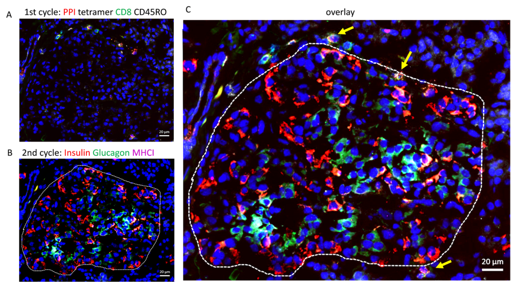
As the slide scanner allows the detection of up to five targets in a single staining, we used a multiplex immunofluorescence staining technique that includes staining cycles using the same tissue section. Briefly, after image acquisition of the first staining, tissue sections were treated chemically to inactive the fluorophores. Then, the same tissue section was re-stained and scanned again.


A) Immunofluorescence images of a pancreas section from a donor with type 1 diabetes stained with HLA-class I tetramer (PPI15-24, red), CD8 (green), the memory marker CD45RO (white) and a nuclear marker (Hoechst, blue) to identify PPI-specific CD8 T cells. Images were taken with the Axio Scan.Z1 slide scanner. Then, after chemical fluorophore inactivation, B) the same tissue section was re-stained for insulin (green), glucagon (red), and MHC class I (magenta) and images were taken with the Axio Scan.Z1 slide scanner. C) The Image shows an overlay of all 7 markers. The white dashed line indicates the islet. The yellow arrows show PPI-specific CD8+ T cells close to the islet. We thank Drs. Sara McArdle (Scientific Associate and Chan Zuckerberg Imaging Scientist) and Zbigniew Mikulski (Director of Microscopy Core, La Jolla Institute for Immunology) for their help and support with image acquisition.
Thanks to the slide scanner technology, we were able to quantify and localize autoreactive CD8 T cells as well as gain critical insight into their distribution in the whole pancreatic tissue section.
Learn More
Read the full article: “The healthy exocrine pancreas contains preproinsulin-specific CD8 T cells that attack islets in type 1 diabetes“
Learn about the ZEISS Axio Scan.Z1 slide scanner, the technology that was used in this publication.


