Sep 1, 2020
Nikon Instruments Inc. is pleased to announce the release of NIS-Elements Clarify.ai, a new addition to Nikon’s growing suite of artificial intelligence (AI) tools for microscopy. Clarify.ai uses artificial intelligence to automatically remove blur from widefield fluorescence microscope images.
Clarify.ai utilizes new Nikon technologies executed on graphic processing units (GPUs) to leverage fast and efficient clarity in images normally corrupted by blur due to out-of-focus light.
Clarify.ai leverages deep learning, a type of artificial intelligence. Clarify.ai is pre-trained to recognize fluorescence signal emitted from out-of-focus planes and can computationally remove this haze component from the image, resulting in significantly improved signal-to-noise (S/N) ratio.
Main Features
1. AI algorithm removes out of focus fluorescent signal
In widefield fluorescence microscopy, image clarity can be impaired by out-of-focus signal emitted from above and below the focal plane. Traditionally, deconvolution is used to reassign out-of-focus light, or additional hardware devices such as confocal pinholes used to reject this light.
Clarify.ai utilizes modern artificial intelligence deep learning technology to recognize out-of-focus light, and can automatically remove this component from widefield fluorescence images.
This new AI-based module enables high-contrast results from any widefield fluorescent sample or acquisition magnification. It can also easily manage removal of heterogeneous scatter while preserving intensity linearity of the underlying data.
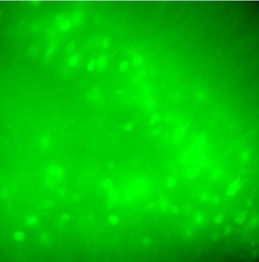 Widefield fluorescence image of brain slice, 40x Widefield fluorescence image of brain slice, 40x | 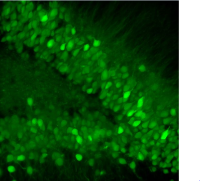 Clarify.ai result image Clarify.ai result image |
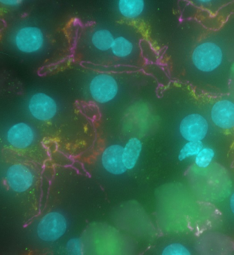 Tissue section, 60x Tissue section, 60x | 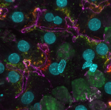 Clarify.ai result Clarify.ai result |
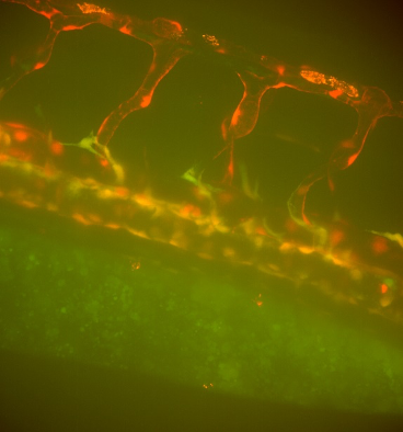 Widefield image of Zebrafish vasculature, 20x Widefield image of Zebrafish vasculature, 20x | 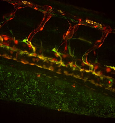 Clarify.ai result image Clarify.ai result image |
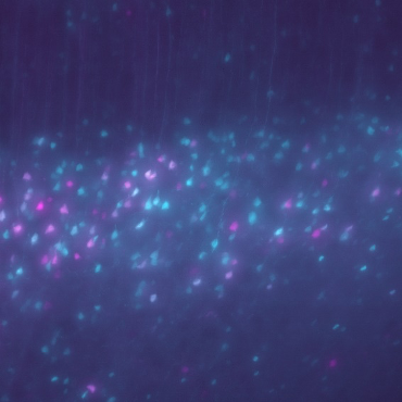 Widefield brain slice image, 40x Widefield brain slice image, 40x | 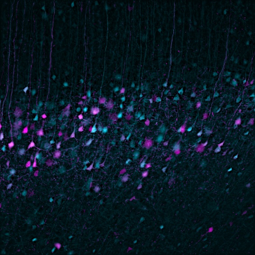 Clarify.ai result Clarify.ai result |
2. Simple execution
Clarify.ai can be used on any widefield 2D or 3D data set obtained from any microscope system, detector, or magnification, without the need for AI training or introduction of bias from complicated user-settings: it has been pre-trained to recognize and remove just the out-of-focus contributions from images and preserve the original details with one click.
3. Applicable to all widefield data sets
No special hardware configuration is required for acquiring images suitable for Clarify.ai; any fluorescence widefield images obtained from any detector can be used: including cell cultures, model organisms, tissue sections/slices, or other samples.
Widefield microscopy generally enables fast acquisition speeds and low system costs, and is readily applicable to imaging live specimens. Applying Clarify.ai to widefield data sets enables high signal-to-noise ratio results with high contrast while still maintaining the benefits of widefield microscopy.
Computation is fast and can be applied at the experiment time, or later – it can even be applied to archived data for improvement of contrast.
To learn more about Clarify.ai, click here.


