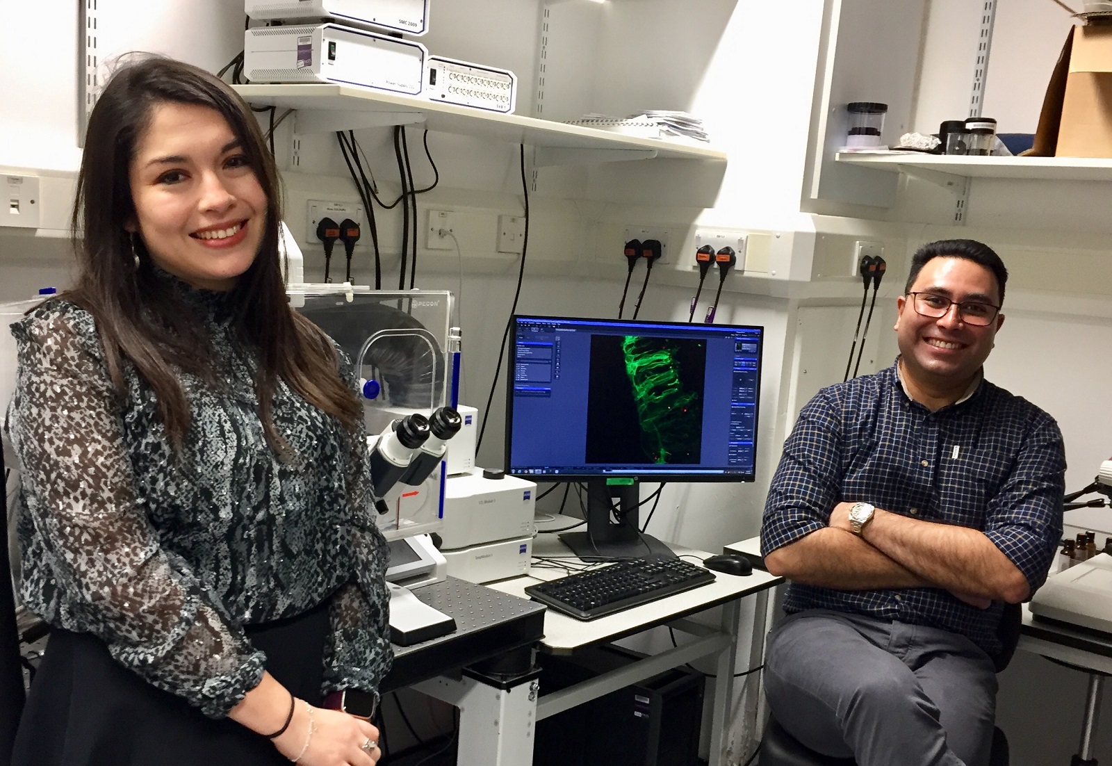
Dr. Raman Das is an independent research fellow at the University of Manchester (United Kingdom). Using the chick embryo as a model organism, Dr. Das studies how cells develop into neurons and create the vertebrate central nervous system. In their recent publication, Dr. Das and his postdoctoral research associate, Dr. Gabriela Toro, used wide field live cell imaging and Airyscan superresolution live cell imaging to further elucidate the mechanics of how these cells migrate.
We interviewed Dr. Das to learn more about his and Dr. Toro’s recent work.
What are your research goals?
The overarching aim of my research group is to investigate the cell behaviour underlying neuronal differentiation in the context of vertebrate embryonic development. This is a fundamentally important process that generates the diverse array of neuronal subtypes which are ultimately required for the formation of functional neural circuitry.
Cells differentiating into neurons in the developing chick spinal cord. Cell membranes labelled with GFP-GPI (green). 50um z-stacks were acquired every 10 minutes over 17 hours with Airyscan.
Although the molecular cascades that drive neuronal differentiation and specification of distinct neuronal sub-types are well understood, we still know very little about how these molecular cascades are translated into the fascinating cell behaviour that drives this critically important process.
To address this, we have developed a unique assay to image cell behaviour during neurogenesis in the context of developing tissues over long periods of time, thus enabling studies at the interface of cell and developmental biology.
What did you discover in your recent publication?
This publication builds on our previous discovery of the regulated process of apical abscission in which newborn neurons in the developing spinal cord cut off their roots as they migrate to their new residence.
This results in loss of their primary cilium, which is needed to process extracellular signals.
In this paper, we have discovered that these newborn neurons rapidly re-assemble a new primary cilium, through a process we call primary cilium remodeling.
A differentiating neuron delaminating from the spinal cord neuroepithelium. This cell sheds its primary cilium (Arl13b-TagRFP, red) but retains a short Arl13b+ particle which will act as the base for reassembly of a new primary cilium. Images are 50um z-stacks acquired every 10 minutes for 3 hours and 10 minutes using wide field microscopy and deconvolved with ZEISS ZEN software.
Our exciting findings have revealed that the plasticity of the primary cilium can be exploited by newborn neurons to drastically modify how they respond to signaling cues from the extracellular environment. This facilitates the vast morphological changes characteristic of axon extension and is therefore essential for normal neuronal differentiation.
How did live cell imaging support your research?
Traditionally, live cell imaging has been technically challenging as long-term imaging of embryonic tissues has been limited by tissue viability and the phototoxic effects of live imaging using fluorescent markers. We overcome these issues by using a novel embryonic slice culture assay in combination with high-end widefield deconvolution imaging, which facilitates live imaging of cell behaviour over several days. The use of widefield imaging is particularly crucial here as it minimises phototoxic effects and, in combination with the highly sensitive sCMOS cameras we use, facilitates imaging of small and dim objects using very short exposure times.
This live-tissue imaging approach has enabled us to image how cells undergoing apical abscission inherit a small portion of the original primary cilium, which then acts as the base for the reassembly of a remodelled primary cilium that is molecularly distinct from the original primary cilium. Using chromophore-assisted light inactivation (CALI), we were then able to disrupt primary cilium remodelling, which resulted in cells being unable to extend a nascent axon. Furthermore, disrupting the remodelled primary cilium in cells that were already extending axons resulted in a dramatic collapse of the extending axon, indicating that primary cilium remodelling is essential for initiation and maintenance of axon extension.
A differentiating neuron undergoing axon extension. The cell membrane is labelled with mKate2-GPI (red) and primary cilium with Arl13b-TagRFP (red). IFT88-mNeonGreen was used to visualize intraflagellar trafficking in the neuronal primary cilium. Images are 50um z-stacks acquired every 10 seconds for 2 minutes using wide field microscopy and deconvolved with ZEISS ZEN software.
Finally, we used our live imaging approach in combination with fixed-tissue imaging to demonstrate that primary cilium remodeling results in a switch from canonical to non-canonical Shh signal transduction, which directs the process of axon extension.
Learn More
Read the full article “Primary cilium remodeling mediates a cell signaling switch in differentiating neurons” Link
Learn about ZEISS solutions for wide field live cell imaging and superresolution with ZEISS Airyscan.


