 August 18, 2010 (Peabody, Mass.) — The ability to examine biological samples with both electron microscopy and light microscopy using a single instrument and switching between the two with a single mouse click is garnering top honors for the JEOL ClairScope.
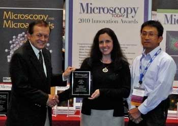
In August, the editors of Microscopy Today magazine selected the ClairScope for the MT-10 award, which recognizes ten microscopy innovations that make imaging and analysis more powerful, more flexible, more productive, and easier to accomplish. The award was given to JEOL during M&M 2010, the Microscopy & Microanalysis conference held in Portland, Oregon.
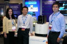
The ClairScope is a truly correlative microscope that combines the power of electron microscopy with light microscopy. Researchers can perform routine fluorescent imaging then examine the specimen at high resolution, high magnification without changing instruments or sample position. The wide-field light microscope with emersion lens is co-axially aligned with the atomospheric SEM column.
This complementary technique employs a unique specimen dish with an ultrathin film window that allows electron beam transmission while the sample is open to atmospheric pressure. Unlike  other SEM techniques, reagents, drugs, and other substances can be added to the sample in order to perform experiments and observe reactions in both liquid and gas environments in real time, typically only possible with the light microscope.
Sample volumes can be as high as 10 mL. The sample holder is compatible with either cell cultures or a wide range of materials (liquids, gels, solids, etc.). Dynamic phenomena such as crystallization, drying processes, and electrochemical reactions (sample holder with electrodes) can be followed in real time. Biological materials can be imaged without the lengthy pretreatment (dehydration, fixation, coating, etc.) necessary in conventional SEMs.
“There are so many new applications for high resolution microscopy as a result of this innovation,” said Donna Guarrera, Asst. Director of the Scanning Microscopy Division at JEOL USA. “Biologists can observe biological processes such as platelet generation, distribution of sugar chains, and microbe growth. Materials scientists will be able to observe and record crystallization, electrochemical reactions, emulsion technology, self-assemblies, and dendrite growth as they occur.”
Also acknowledging the significance of this new instrument, the R&D 100 Award, known as the “Oscar of Innovation,” was bestowed on the JEOL ClairScope on July 8 by the editors of R&D Magazine.
The first JEOL ClairScope to be in operation in the United States is being installed at Northwestern University’s Biological Imaging Facility where it will be used for demonstrations and applications development.
https://www.jeolusa.com/NEWS-EVENTS/Press-Releases/PostId/101/JEOL-Correlative-Microscope-Wins-MT-10-Award


