Neuroscientists investigate brain connections using zoom and confocal microscopy to better understand how specific neural circuits generate behavior in normal animals and in disease models.


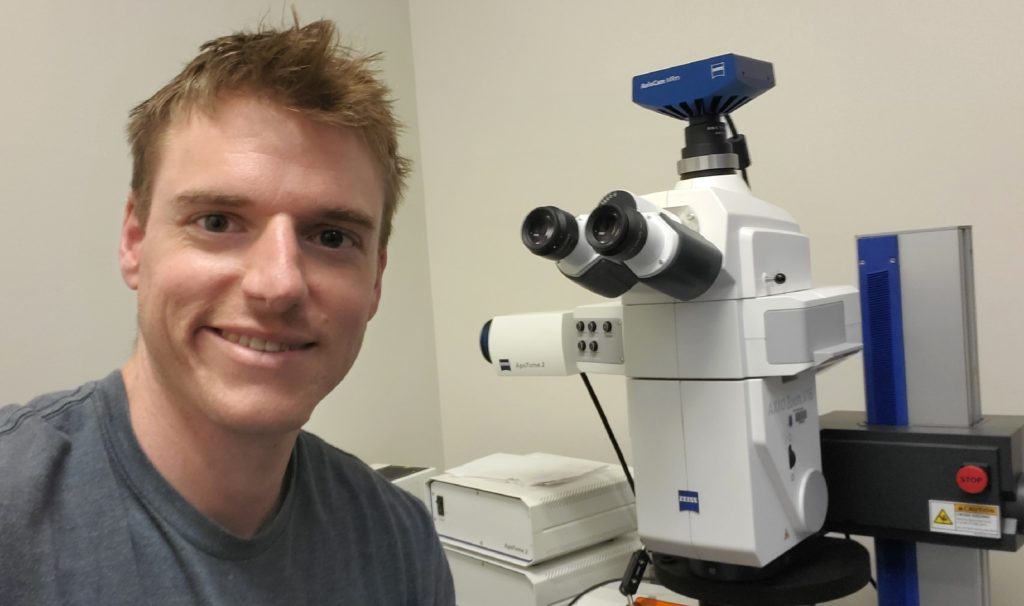
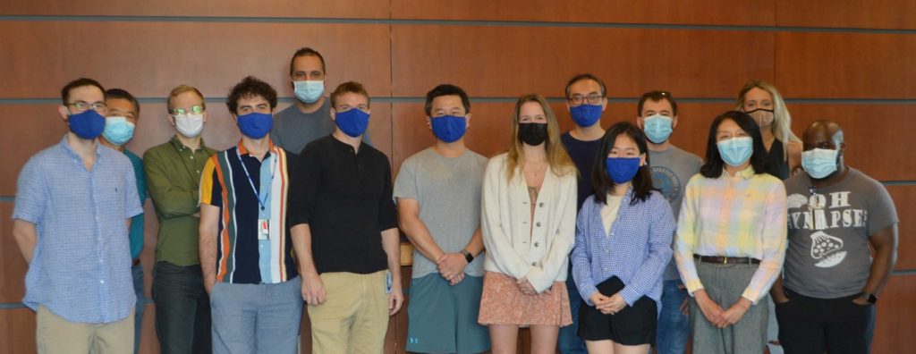
Dr. Ryan Hughes (top image) and Dr. Henry Yin (lower image, center, gray shirt) with his lab, including Dr. Hughes, who is directly to his right.
If you are warm and sitting in your home, you likely have many options to make yourself more comfortable. You could remove your sweater, lower your thermostat, open a window or walk outside.
This was the example Dr. Ryan Hughes, a postdoctoral researcher in Dr. Henry Yin’s laboratory at Duke University (USA), provided when asked to explain the phrase “achieving a fixed end with variable means.” He explained that neuroscientists still do not know how the brain produces behavior and do not have a successful model that can replicate behavior where fixed ends are achieved with variable means. Our lack of understanding is so large, he explained, that we cannot even model the most basic behaviors of the simplest of organisms, such as that of the fly or the worm.
The Yin lab uses an integrative approach to study of nervous system, combining electrophysiology, calcium imaging, optogenetics, behavioral analysis and anatomical tracing.
We spoke with Dr. Hughes about his work on a recent article from the Yin lab which used zoom microscopy and confocal microscopy to better understand some specific connections within the brain that could be targeted to better alleviate Parkinsonian symptoms.
What research question were you focusing on in this article?
In Parkinson’s disease, dopamine neurons degenerate and die resulting in bradykinesia (slowness of movement), akinesa (lack of movement) and tremors. The most successful treatments for Parkinson’s disease are L-Dopa (a dopamine precursor that supplies the brain with more dopamine) and deep brain stimulation (DBS) in the subthalamic nucleus (STN); however, these still do not effectively cure the disease. In addition, we do not know why stimulating the STN alleviates Parkinsonian symptoms.


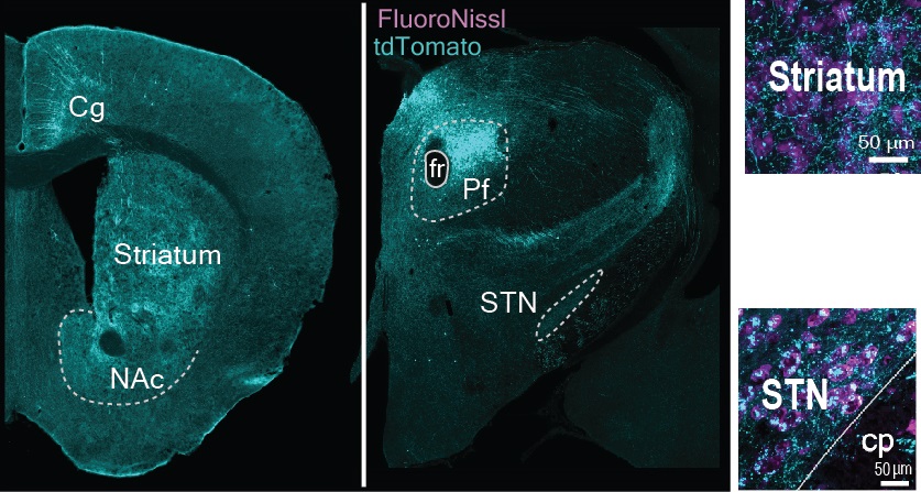
CAV-2, a fluorescent tracer labeled with tdTomato, was injected into the parafascicular (Pf) nucleus. This allows visualization of neurons that are both travelling to the Pf nucleus in the forward (efferent) or backward directions (afferent). FluoroNissl stains all cell bodies; this shows all the neurons regardless of their type. Image collected using a ZEISS confocal microscope.
Traditional DBS methods use non-selective electrical stimulation, which could activate and inactivate multiple cell types and neural pathways. In our study, we performed selective activation of the parafascicular (Pf) nucleus and its projections to the STN.
How did microscopy contribute to your findings?
We first investigated the Pf’s connections with the striatum and, to our surprise, we found that it did not have much impact on movement or alleviating Parkinsonian symptoms.
Microscopy then played an extremely important role. After we did not get any significant effects from stimulating the Pf projections to the striatum, we conducted several anatomical experiments with both retrograde and anterograde fluorescent tracers. This allowed us to visualize the input-output connectivity of the Pf and pinpoint the terminals in other basal ganglia nuclei to which the Pf projects. We used both zoom microscopy for lower resolution images and confocal microscopy for higher resolution. Using these microscopes, we were able to elucidate the circuits of the Pf, and in particular the Pf-STN circuits, with much higher precision and cell-type specificity than what had ever been done previously.
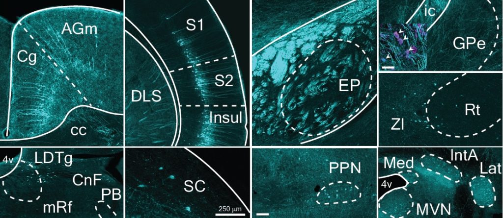
Examples of the output and input projections from the Pf (blue). By using a trans-synaptic anterograde tracer (it jumps the synapses between neurons) we were able to visualize their di-synaptic projections through the STN (green). Images acquired with a ZEISS confocal microscope.
We hypothesized that the Pf projections to the STN could be responsible for action initiation and contribute to the beneficial effects of traditional STN DBS. When we selectively stimulated this pathway, we could alleviate numerous pathological symptoms characteristic of Parkinson’s disease.
Learn More
Read the full article, “Thalamic projections to the subthalamic nucleus contribute to movement initiation and rescue of parkinsonian symptoms” Link
Learn about ZEISS solutions for zoom microscopy and confocal imaging.
Read Next


