From scanning electron microscopy data to tactile teaching tools
In 2017, Dr. Alex Ball, head of Imaging and Analysis at the Natural History Museum London (NHM), approached ZEISS with an intriguing idea. What if one could take 3D microscopy data sets and create tangible objects using 3D printers? This would create a physical teaching object for students with visual impairments. We previously wrote about the start of this project which used data from scanning electron microscopes.
ZEISS partnered with Dr. Ball and agreed to support a CASE award for a PhD project. The project is now in its third year and is a collaboration with NHM, University College London and ZEISS. We connected with Dr. Ball and he was able to share some of the new and improved 3D teaching tools he and his PhD student, Kate Burton, are designing.
A closer look atha common objects
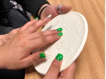
Dr. Ball and Mrs. Burton worked to create 3D models of common objects that we all have felt in everyday life: coins, sand and sugar grains. By printing these objects at different magnifications, one can experience the principle of magnification through tactile methods.
Images: Coins were 3D printed at 5x, 10x, 20x and 50x magnification. Sand and sugar grains were imaged with scanning electron microscopy (SEM) and 3D printed at 50x magnification. The coin and a grain of sugar, both 3D printed at 50x magnification, are compared side by side.
The feedback they have acquired from workshops of visually impaired students are very positive. Touching these magnified objects helps them to understand the fine structures that they feel in their fingertips.

One of them said they did not realize that there was a face on the coins until they touched and felt the magnified 3D version of it.
Entomology through touch
In 2017, Dr. Ball and colleagues had published work first detailing how to collect data of small objects for 3D model printing. In this paper, they had collected data for various insect heads using scanning electron microscopy (SEM) and then using that data to create magnified, 3D models.
Insect heads of the damselfly, butterfly and housefly created from scanning electron microscopy (SEM) data and 3D printed at 25x magnification.
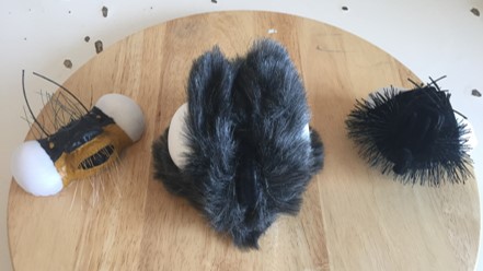
More recently, Mrs. Burton and Dr. Ball supervised a Masters student, Emma Martin, who is studying Disability Design and Innovation at UCL, who redesigned the original models to create more realistic teaching objects. The challenge was not only to accurately model the shape, but also the feel of the specimen, such as the fine hairs on the butterfly as well as the hard and soft parts of the creatures. The goal was to create a process that could be easily replicated by a school group as part of a STEM project. Ms. Martin’s thesis was awarded a Distinction for the new models she created along with the accompanying teaching course.


Newly designed damselfly, butterfly and housefly heads.
What’s making me sneeze?
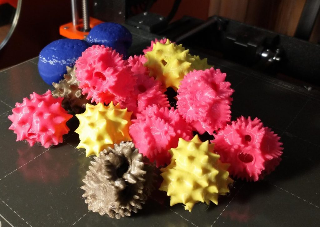
Dr. Ball is now also working with Oliver Wilson, a PhD student at York University and creator of the 3D Pollen Project. His goal is to produce 3D-printed models from high-quality confocal microscopy scans of pollen grains and to make them all freely available for outreach, education and research. While their goal is to create new data of pollen grains, Dr. Ball is currently 3D printing pollen grains from Mr. Wilson’s publicly available data.

Pollen grains 3D printed by Dr. Alex Ball using confocal microscopy data from Oliver Wilson’s 3D Pollen Project.
A portable museum at your fingertips
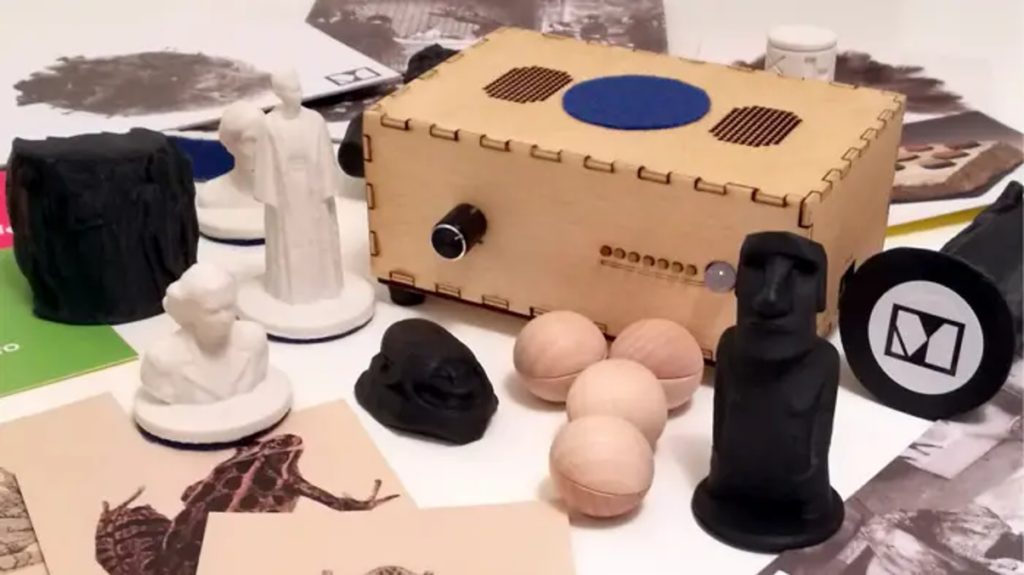
Dr. Ball and Mrs. Burton are working with the creators of “Museum in a Box” to include their 3D models in this novel concept for educational outreach.
Museum in a Box is a portable, interactive exhibit using either 3D models or cards containing an embedded RFID chip and a small box containing a programmable chip-reader equipped with a speaker. By placing the model on the box, playback of an audio file is triggered which provides information about the model.

Museum in a Box
Dr. Ball and Mrs. Burton hope to incorporate their 3D models into a “Museum in a Box” and use in outreach to students.
Waiting out the pandemic
As Dr. Ball and Mrs. Burton wait out the pandemic, one new set of models they are working on is a 3D print of SARS-CoV-2, the virus which causes COVID-19.
Data have been made available from the Coronavirus Structural Task Force (University of Hamburg) to 3D print models of the coronavirus based on the most up-to-date scientific models available. Prints are at 1,000,000x scale and are suitable for all types of 3D printers.
Coronavirus Structural Task Force
They are looking forward to adding these as another teaching object to their growing gallery of 3D models.
Learn more
Read the paper where Dr. Ball first detailed how to collect data of small objects for 3D model printing using scanning electron microscopy (SEM).


