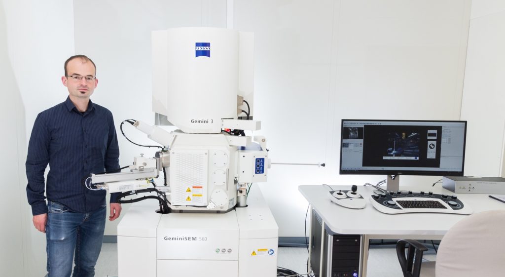
The Center for Applied Quantum Technology (ZAQuant) receives the world’s first ZEISS GeminiSEM 560 installation
The precise measurement of physical quantities is a driving force for almost all technical progress. The key mission at the Center for Applied Quantum Technology (ZAQuant) at the University of Stuttgart (Germany) is the development of novel quantum sensors for improved sensitivity, specificity and energy efficiency.

Dr. Mario Hentschel with the newly installed ZEISS Gemini 560 FE-SEM.
Their research will now be enhanced by the addition of a ZEISS GeminiSEM 560 FE-SEM. This tool is one of the new models of ZEISS electron/ion microscopes launched in November. ZAQuant has received the first installation of this new system.
We spoke with Dr. Mario Hentschel, Head of Cleanroom and Nanostructuring Facilities, about their use of field emission scanning electron microscopy, their experience with GeminiSEM and some example images they have acquired with their new tool.
Tell us about your job at ZAQuant.
I’m a mixture of a researcher and an experimental officer. I look after the clean room by managing the space and keeping it functional. I do some training, but I also use the equipment a lot myself to manufacture the micro- and nano-structured devices we need for our experiments. I work on characterization of the structures we produce, for which I utilize the new GeminiSEM.
How does Field Emission Scanning Electron Microscopy (FE-SEM) support your work?
It is important to see our devices on a nanometer scale.
FE-SEM is used to control the nano- and microstructuring process. We use a clean room which is equipped with various instruments for nanoscale structuring. Quality needs to be carefully controlled in these small dimensions.
We also use FE-SEM to characterize the microstructures and correlate with their optical properties. We use EDS to identify impurities or other defects. We need precise measurements of the dimensions of the structures. These need to be measured precisely with the help of the microscope.
Electrochemically grown single crystalline gold. Simon Mangold and Bettina Frank. 4th Physics Institute, University of Stuttgart. Electrochemical methods allow the synthesis of mono- and polycrystalline gold structures. High resolution imaging is critical to investigate the growth mechanism and optimize and tune the growth. Apart from monocrystalline structures, which exhibit perfect hexagonal or triangular shapes and smooth surfaces, polycrystalline structures with irregular shapes are observed. For more information: Science Advances 3, e1700721 (2017).
Were there any particular features of GeminiSEM that are useful for your work?
I was very impressed with the flexibility of the system. There are various detectors available to image and highlight topography, material contrast, edges on the surface and all the different features of our samples. The system can image at both low and very high magnification. Imaging at the highest magnification is important, but we have to deal with very different materials ranging from metal to ceramic and sometimes even organic molecules. It was important to be able to image without suffering from charging.

Single crystalline gold nano bipyramids, tips capped with palladium Qi Ai, 4th Physics Institute, University of Stuttgart. Sample prepared by the group of J. Wang, Department of Physics and Shenzhen Research Institute, The Chinese University of Hong Kong. Advanced wet chemical synthesis methods allow for the creation of complex colloidal nanostructures. The SEM image shows bipyramidal gold nanorods, which are additionally dressed with palladium dots at the two tips. This allows the use of these structures as local plasmonic sensors and reporters. Imaging of these rods is often hindered by the remaining surfactants and other chemical compounds which are prone to strong charging effects. To investigate the spatial distribution of the two materials and therefore optimize the synthesis, EDX is used. The Au signal reveals the bipyramidal shape of the Au crystals below the Pd overcoat. For more information: H. K. Yip et al., Advanced Optical Materials 5, 1700740 (2017)
What have been your experiences so far with GeminiSEM?
We are absolutely amazed by our new SEM.
The stability of the system is excellent, much better as compared to our previous tool.
The STEM capability has proven both insightful as well as highly convenient. For daily characterization, a dedicated TEM is no longer necessary. We were surprised that we could resolve DNA substructure, even in STEM, without the need for TEM.
GeminiSEM offers a very high degree of freedom, both regarding the compatibility with many different sample structures, as well as regarding the choice of detector and detector information.

Magnesium Crystalline Films. Active plasmonic and nanophotonic systems require switchable materials with extreme material contrast, short switching times, and negligible degradation. On the quest for these supreme properties, an in-depth understanding of the nanoscopic processes is essential. Magnesium, which has proven to be an excellent hydrogen storage material, also features a prominent metallic magnesium (Mg) to dielectric magnesium hydride (MgH2) phase transition. Understanding the diffusion behavior in these crystalline films is highly important in order to overcome the limited diffusion coefficients and has substantial impact on the further design, development, and analysis of switchable phase transition as well as hydrogen storage and generation materials. The images show (from left to right): Tilted SE SEM, BF STEM, BF STEM with simultaneous inLens. For more information: J. Karst et al., Science Advances 6, eaaz0566 (2020).
What are you working on now?
In 2021, we are going to move into the newly built home for the center for ZAQuant here at the University of Stuttgart. There will be a range of users and PIs with different needs. We needed enabling technology for their research and felt that this instrument can provide it in a very flexible way. I expect changes with new researchers coming on board and we are now prepared to support them with instrumentation that meets their needs.
Electron-driven Photon Sieves, 4th Physics Institute, University of Stuttgart, Institute for Experimental and Applied Physics, Christian Albrechts University, Kiel. Complex hole patterns are structured via FIB lithography in gold films on top of a TEM membrane. The interaction of electron beams with these structures results in the emission of tailorable complex light fields. Inspection of the structures is crucial for the pattern optimization. For more information: N. Talebi et al., Nature Communications 10, 599 (2019) and N. van Nielen et al., Nano Letters 20, 5975 (2020).
Learn More
Additional research highlights from ZAQuant:
- A molecular quantum spin network controlled by a single qubit. Link
- Nanoengineered diamond waveguide as a robust bright platform for nanomagnetometry using shallow nitrogen vacancy centers. Link
- Coherent control of single spins in silicon carbide at room temperature. Link
Get more information on the new ZEISS GeminiSEM FE-SEM family.


