We previously spoke with Dr. Parmesh Gajjar, a research associate at the University of Manchester (United Kingdom), about his work with X-ray microscopy to study the microstructure of dry powder inhaler medicines. At that time, he was preparing for a presentation where he showed he was able to visualize drug-excipient interactions within a formulation and observe that different crystal facets have different behavior. You can now find this work published here.
We checked in with Dr. Gajjar to find out what he’s been working on recently. He shared with us a new publication from the journal CrystEngComm using X-ray microscopy to investigate crystalline powder bed structure. We chatted with him a bit about this latest work.
Why study crystalline powder bed structure?
The vast majority of pharmaceutical formulations involve polycrystalline materials in some form. The interactions between different crystals can be important for the behavior of the formulation and necessary to assess manufacturability and performance. Surprisingly, there is an absence of methods and techniques for 3D crystallographic information for organic materials and a clear need for techniques that provide non-destructive granular information of organic materials in powders.
How did you use X-ray microscopy to study organic materials in powders?
We had seen that LabDCT, an analytical module of the ZEISS Xradia Versa X-ray microscope, had been successfully used to study 3D crystallographic information, including grain centroid, orientation and shape, for metals, metalloids and minerals. We wanted to test if this technique could be used for organic materials.
To do this, we started with the simple organic crystal hexamine. This has a cubic crystal structure, i.e. its crystal shows the most symmetry and so it should be the easiest for LabDCT.
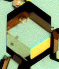
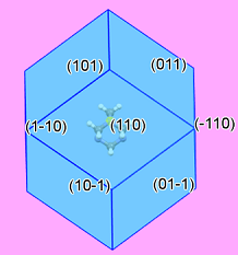
Image: Left: Optical microscopy image of hexamine showing the rhombic dodecahedron crystal morphology. Right: Predicted crystal morphology view looking down. Images courtesy of Hien Nguyen, The University of Leeds, and reproduced from Gajjar et al (2021) under a CC-BY license.


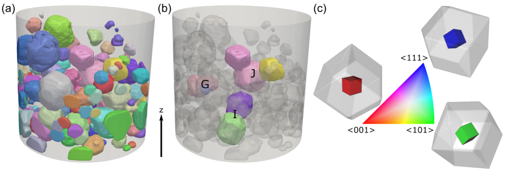
In the paper, we show how LabDCT does indeed work for organic materials and we are able to experimentally prove the crystal morphology as well as reveal the nature of interactions between individual crystals within the powder bed.

Image: (a) 3D visualizations of all the different crystals within the powder identified by LabDCT, each colored according to their orientation. (b) Several groups of crystals, highlighting crystal-crystal interactions. (c) Orientational color bar, with sketches showing what each orientation means in terms of a hexamine crystal. Image reproduced from Gajjar et al (2021) under a CC-BY license.
These results have significance for the pharmaceutical industry and, more broadly, fine chemical industries, where a firm knowledge of the chemical basis behind powder bed structuring is of utmost importance for manufacturing and performance.
Learn more
Read the article Gajjar et al. (2021) “Crystallographic tomography and molecular modelling of structured organic polycrystalline powders” CrystEngComm, DOI:10.1039/D0CE01712D
Learn about the ZEISS technology mentioned in this article including the ZEISS Xradia Versa X-ray microscope and the analytical module LabDCT.
Read Next


