Research highlights use of ZEISS MultiSEM and ZEISS Sigma electron microscopes
Globally, macular degeneration of the retina is a leading cause of legal blindness; however, little is known about its cause. A recent article by Dr. Charles Zucker, Staff Scientist at Harvard University, and colleagues describes electron microscopic observations from the retina of a woman afflicted with a rare form of macular degeneration called macular telangiectasia (MacTel).

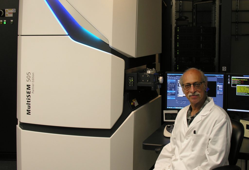
Dr. Charles Zucker, Staff Scientist, Harvard University
For the purposes of learning about pathological mechanisms, where ultimately many samples will need to be analyzed, it is not practical, nor advantageous, to image the entire volume – a more ‘traditional’ connectomics approach.
Zucker refers to the approach used in this study as “targeted high-throughput connectomics.”
The actual block of tissue examined covers a retinal area of about 2 mm x 3 mm, with a thickness in the order of one third of a mm. Slicing the volume into approximately 30 nm thin sections resulted in about 10,000 serial sections, collected with an ATUMtome onto plastic tape. This process is outlined in this video:
Lower resolution overview images of every 100th section were acquired with a ZEISS Sigma FE-SEM electron microscope. This intermediate step was used for guiding the imaging and locating the actual regions of interest (ROIs). Selected ROIs were finally acquired at high resolution and at high speed utilizing a ZEISS MultiSEM electron microscope.
The major observations made were specific changes in mitochondrial structure in all retinal cell types within and outside the MacTel zone. They also identified an abrupt boundary of the MacTel zone that is coincident with the loss of Müller cells and macular pigment. Since Müller cells synthesize serine for uptake by retinal neurons, they propose that a deficiency of serine, required for mitochondrial function, causes mitochondrial changes that underlie MacTel development. Variations in mitochondrial pathology and stages of degradation are shown in a supplementary movie of an aligned stack of images.


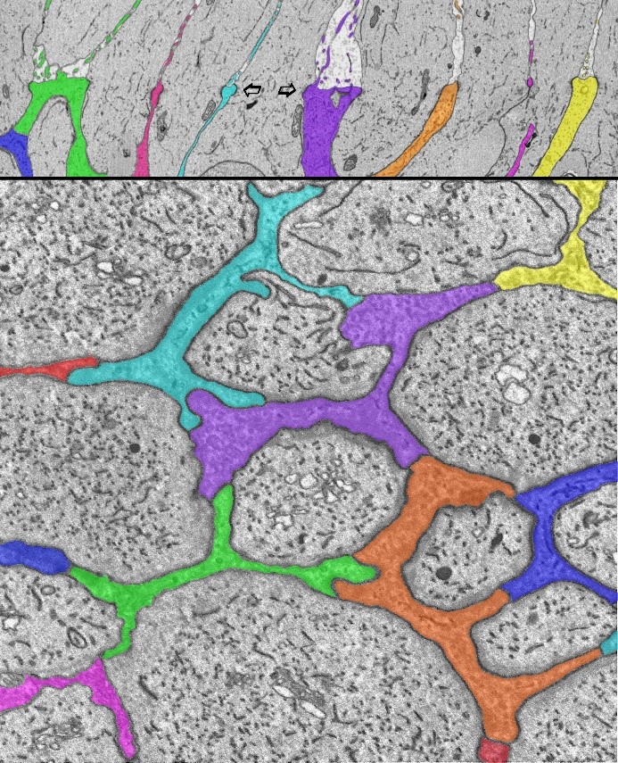
Seen here at the outer limiting membrane of the human central retina, Müller glial cell processes (colored) surround the inner segments of the photoreceptor cells, and interdigitate with them (arrows) and with each other. This forms a specialized structure that constitutes part of the blood-retinal (brain) barrier. High-throughput serial-section electron microscopy reveals the fine details of that glial/neuronal relationship oriented radially (top, arrows) and horizontally (bottom).
The upper image on the left is from an eye with pathology in the central macula region of the retina – but taken from a more peripheral region that is relatively normal and served as an internal control.
The bottom image is from the central macula region (fovea) from another human eye without known disease or pathology. It is from the same z-depth (layer) of the retina that is shown going across the middle of the top image.
Both images were collected by a ZEISS Sigma FE-SEM and used to guide the selection of ROIs for more extensive imaging within the highly pathological region of the tissue with the ZEISS MultiSEM electron microscope.
Zucker explained that this illustrates how the single-beam ZEISS Sigma and the multiple-beam ZEISS MultiSEM complement each other in obtaining an efficient workflow.
Learn More
Read the full article: A connectomics approach to understanding a retinal disease
Learn about the ZEISS technologies used:


