Researchers use scanning electron microscopy and energy dispersive spectroscopy to better understand the materials used in vital pulp therapy

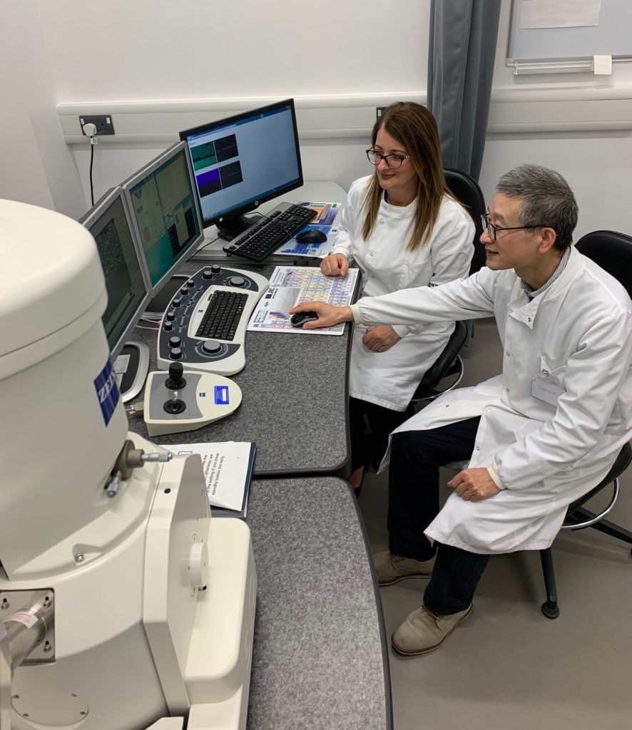
Dr. Josette Camilleri, the academic leading the research on hydraulic cements, at the University of Birmingham and Dr. Jianguo Liu who runs the scanning electron testing (SEM) facility at the School of Dentistry during one of the sessions using a ZEISS SEM for microstructural and elemental analysis
If you are not a dental care professional, you likely have not heard of mineral trioxide aggregate (MTA). It is a hydraulic calcium cement used in dentistry and one of the main subjects of research of Dr. Josette Camilleri, a clinical academic at the University of Birmingham School of Dentistry in the United Kingdom.
Hydraulic calcium silicate cements are unique in dentistry as they set and develop their properties in a wet environment. These cements have been shown to increase clinical success rates in vital pulp therapy – a restorative dental procedure that aims to treat teeth with compromised dental pulp. This is due to the release of calcium and hydroxyl ions from the cement which have antimicrobial properties and also promote tissue healing.
In a new article in Scientific Reports, Dr. Camilleri and her colleagues investigate a series of commercial and prototype materials used for vital pulp therapy to better understand how differences in their composition affects their biological and antimicrobial properties. One of the tools she uses in this publication is scanning electron microscopy for microstructural analysis and energy-dispersive spectroscopy for elemental analysis of these cements.

The team of researchers involved in the publication, from left: Andreas Koutroulis (post graduate research student), Dr. Sarah Kuehne (microbiologist) and Prof. Paul Cooper (oral biologist)
Could you provide further background on MTA? Why is there a need to investigate new cements?
MTA has a very interesting history in dentistry – particularly in Endodontology – which is a dental specialty concerned with the biology and pathology of the dental pulp and periapical tissues.

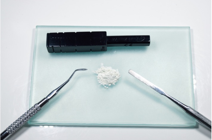
MTA being prepared by mixing the powder and liquid on a glass slab. The MTA block facilitates the delivery to the operating site.
MTA is composed of Portland cement, which is used in the construction industry, and a radiopacifier bismuth oxide, which allows the visualization of the material on a radiograph. Although the use of a construction cement as a biomaterial was revolutionary, construction cements include heavy metal ions and aluminium, which are not indicated for clinical use. Furthermore, the bismuth oxide present in a number of hydraulic cements leads to tooth discoloration. There are also other disadvantages with using the first generation of this material type such as the dispensing, manual mixing and difficult clinical handling.
Scanning electron microscopy and EDS were some of the tools that you used in this paper. Could you elaborate on the use of these technologies?
Scanning electron microscopy and energy dispersive spectroscopy are essential tools for material characterization. They are the backbone on which further material analyses can be made. In this paper, we analyzed polished sections of all the materials after 28 days in physiological solution. We could determine the material ultrastructure and the energy dispersive spectroscopic analyses enabled us to complete precisely the phase analyses by X-ray diffraction. The phase identification is not possible without prior energy dispersive spectroscopic analysis, particularly with commercial products.

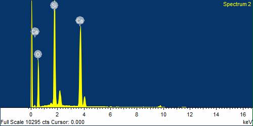
Back scatter scanning electron micrographs of Bio-C Pulpo (Angelus Londrina Brazil), a clinically available hydraulic calcium silicate cement, and one of the prototype materials being tested at the School of Dentistry, University of Birmingham, to elucidate the effect of additives on the material microstructure; The energy dispersive spectroscopy plot of the main elements found in tricalcium silicate cements
Was there anything you found surprising or particularly interesting when doing this research?
All calcium-based materials used in dentistry are specifically aimed at the biological interaction. The calcium ion release has always been implicated in calcific barrier formation over the dental pulp with the related biological effects. Although dental diseases are all bacteria induced, the antimicrobial characteristics of materials still does not seem to be given enough importance. In fact, antimicrobial properties of materials used for vital pulp therapy are scarcely investigated and the interplay between antimicrobial and biological characteristics even less so. We also wanted to check if the calcium ion effect was dose related. This research clearly underpins the importance of antimicrobial testing of all materials used for vital pulp therapy and the need to develop smart materials with specific dose-related effects targeted at specific responses.
Learn More
Read the full paper “The role of calcium ion release on biocompatibility and antimicrobial properties of hydraulic cements“
Get information on ZEISS scanning electron microscopes
University of Birmingham is a ZEISS labs@location. This title is bestowed to organizations which offer a community of ZEISS customers and technologies, providing in depth knowledge and dedicated services. Find a partner labs@location site to assist you with your research using the link below.


