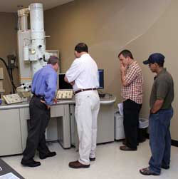
 Peabody, Mass., July 25, 2006 — JEOL USA, the industry-leading supplier of electron microscopes, has installed a model JEM-2100 LaB6 Transmission Electron Microscope (TEM) at Oklahoma State University’s new microscopy service laboratory. The lab, which will serve researchers from both the university and neighboring industry, hosted an open house to introduce their expanded facilities and services, including the cryogenic capabilities of the new TEM.
Professor’s Dream Realized
The new lab is the vision of an OSU professor who established the original microscope service lab in the basement of OSU’s veterinary school nearly 30 years ago. Dr. Charlotte Ownby had recently retired , but returned to become director of the new electron microscopy lab, named Venture I and located in the Oklahoma Technology and Research Park in Stillwater.
Armed with a grant from the National Science Foundation, Dr. Ownby purchased the new Transmission Electron Microscope (TEM) from JEOL USA in Peabody, Massachusetts. “There was good science in our proposal,” Dr. Ownby says. “We were able to show the commitment of the University with a brand new lab and a 50% increase in space.” Through the vision of key people including Steve McKeever, Vice President for Research, the new lab was funded by OSU.
Venture I Lab Serving Industry and Science
The on-campus lab served some 200 users from various colleges, whereas the new lab, less than a mile from campus, will extend its services outside the university clientele. Ownby hopes to attract new businesses and potential clients coming in the research park.
“The University needs a good position to interact with industry, especially high tech. It’s already paid off,” says Ownby. Three new projects were acquired by the full service lab the day after the open house held in May.
New Capabilities with Cryo TEM
The cryogenic capabilities of the new TEM open a whole new frontier to researchers, who can now study frozen samples such as animal tissue, protein structures, vegetables, bacteria, viruses, and a variety of nanomaterials that are preserved in a near-natural state without chemical alteration. This affords a more realistic view of the cellular or atomic structure of the samples which are observed at magnifications up to 1.5 million times.
Current and New Applications
Some of the applications for this new instrument include those of an animal disease diagnostic lab, which uses TEM analysis to determine if a live animal is infected with a virus. A local manufacturer uses the electron microscope services to inspect metal parts used in space missions. For another client, the lab combines x-ray analysis, confocal microscopy, and TEM to determine the toxicity of nano-particles in biological tissues. “It’s odd for an electron microscope lab to have a confocal microscope, but it’s a powerful, more convincing approach because it gives you the bigger picture,” says Ownby. Employing three people, the lab provides results in a timely and cost-effective manner.
The lab will also utilize the new TEM for elemental and x-ray analysis of samples with emphasis of determining the chemical composition in addition to structure. A scanning electron microscope, an atomic force microscope, and the confocal microscope are other key pieces of equipment.
Ownby Renowned Microscopist and Scientist
Dr. Ownby is one of the co-founders of the Oklahoma Microscopy Society, established in 1977, which has 80-100 members, including many from the oil industry and universities. Until retiring from her faculty position at OSU last January, she headed an internationally recognized snake venom research program and was president of the International Society on Toxinology. After graduating from the University of Tennessee’s pre-med program, she was a National Science Foundation research participant and obtained a Master of Science degree in Zoology. She has a Ph.D. in Veterinary Anatomy from Colorado State University.
https://www.jeolusa.com/NEWS-EVENTS/Press-Releases/PostId/37/New-Electron-Microscopy-Lab-Opens-with-Latest-Cryo-Technology-from-JEOL


